Dermatologists managing high-risk patients face a decision that did not exist twenty years ago: how much imaging is enough, and what kind? The proliferation of digital dermoscopy devices and total body photography systems has given clinicians more options than ever. It has also created more confusion about which approach delivers the best clinical outcomes for which patient cohort. This article breaks down the practical differences between spot dermoscopy and total body photography, examines the clinical evidence for each, and offers a framework for deciding which approach, or which combination, is right for a specialist clinic.
- Spot dermoscopy examines individual lesions in detail. Total body photography documents the entire skin surface to detect new or changing lesions over time.
- Total body photography is most clinically valuable for high-risk patients, those with high mole counts, a personal or family history of melanoma, or immunosuppression.
- For most dermatology clinics, the optimal approach combines both: total body photography for baseline documentation and spot dermoscopy for detailed lesion assessment.
- Advanced systems integrate both into a single workflow, reducing consultation time and improving diagnostic accuracy.
Defining the two approaches
Spot dermoscopy
Spot dermoscopy involves examining individual lesions using a dermoscope, either handheld or digital, to assess dermoscopic features such as pigment network, regression structures, vascular patterns, and colour distribution. It is the standard tool for detailed lesion assessment and is used in the vast majority of dermatology consultations involving skin lesion review.
Digital spot dermoscopy adds image capture and storage, allowing the clinician to photograph a lesion and compare it against images from previous appointments. This enables serial monitoring, the practice of tracking a specific lesion over time to detect subtle changes that may indicate malignancy.
Total body photography
Total body photography involves photographing the entire body surface systematically, creating a comprehensive baseline of all visible lesions. The photographs are taken from standardised angles and distances, covering defined body segments. This baseline is then used at future appointments to identify new lesions and lesions that have changed.
Unlike spot dermoscopy, total body photography is not designed to examine individual lesions in microscopic detail. Its value lies in providing a complete map of the skin at a point in time, so that nothing is missed between appointments, including new moles that may not yet be on the clinician’s radar.
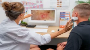
ALT Texts: Dermatologist reviewing skin lesion images with patient using dermoscopy device
When spot dermoscopy is the right tool
Spot dermoscopy is the appropriate primary tool in the following clinical situations: ● Assessment of a specific lesion presenting for the first time with suspicious features ● Follow-up monitoring of a known lesion that has been flagged for ongoing review
- Pre-excision assessment to confirm clinical suspicion before referral or treatment
- General practice skin cancer screening where comprehensive body mapping is not indicated For most outpatient dermatology consultations, spot dermoscopy is sufficient. The limitation is that it is reactive — it assesses lesions the clinician or patient has already identified. It does not systematically survey lesions that neither party has noticed.
When total body photography is the right tool
Total body photography becomes clinically essential when the patient profile shifts to high risk: ● Patients with more than 50 melanocytic naevi, where tracking individual lesions manually is impractical ● Patients with a personal history of melanoma, where the risk of a second primary is significantly elevated ● Patients with a family history of melanoma in first-degree relatives
- Patients on long-term immunosuppressive therapy, who have an elevated skin cancer risk ● Patients with dysplastic naevus syndrome
For these patients, total body photography does something spot dermoscopy cannot: it identifies lesions that are new since the last appointment. A new mole is one of the most clinically significant early indicators of melanoma, but it is only detectable if there is a baseline to compare against. Without total body photography, a new lesion can go unnoticed for months or years.
The case for combining both approaches
In practice, the most effective approach for a specialist dermatology clinic is not to choose between spot dermoscopy and total body photography — it is to use both as part of an integrated workflow.
Total body photography establishes the baseline. It documents what exists at a given point in time. Spot dermoscopy then provides the detailed assessment of individual lesions that are flagged as suspicious or that have changed between appointments.
This combination delivers something neither approach can achieve alone:
- Completeness: no new lesion goes undetected because the entire skin surface has been documented ● Precision: individual lesions of concern are assessed in high-magnification detail
- Continuity: the patient has a comprehensive documented history across every appointment The logistical objection to this approach has historically been time and cost, total body photography required a dedicated room, specialist equipment, and significant staff time. That is no longer the case with current technology.
What current digital imaging systems offer
Advanced systems like the MoleMax HD Pro (molemaxsystems.com/product-molemax-hd-pro/) integrate total body photography and high-definition spot dermoscopy into a single platform. The system uses dual touchscreen monitors for efficient workflow, supports up to 100x optical magnification for detailed lesion assessment, and includes automated total body mapping across 33 preset body segments. A Canon SLR integration provides high-resolution full-body imaging alongside the dermoscopy camera.
Key clinical features of the MoleMax HD Pro that are relevant to the spot versus total body photography decision:
- Total Body Mapping Session: systematic full-body photography with integration of a digital SLR for high-resolution baseline images
- Real Time and Overlay Follow Up: previous and current lesion images displayed side by side or overlaid for immediate comparison
- Thumbnail Monitoring: combines micro image, macro image, and body location simultaneously for quick and efficient follow-up sessions
- Mole Count Module: automatically detects naevi in follow-up images and compares against baseline, extracting size, shape, and brightness features for each lesion
- ABCD Automatic Scoring Module: calculates the ABCD score automatically for each lesion to support consistent, objective risk assessment
- No cloud storage, no subscription fees: patient data stays local within the clinic
For dermatology clinics evaluating integrated digital imaging systems, the MoleMax HD Pro offers a free demonstration at molemaxsystems.com/product-molemax-hd-pro/.
Practical considerations for clinic implementation
When implementing either approach or both, there are several practical factors to consider: Patient selection for total body photography
Not every patient needs total body photography. Establishing clear criteria for which patients are offered total body photography at their initial appointment avoids unnecessary cost while ensuring high-risk patients receive appropriate monitoring. The criteria above — mole count, personal history, family history, immunosuppression, provide a practical framework.
Consultation structure
Integrating total body photography into a consultation requires a dedicated appointment slot. Most clinics schedule total body photography as a separate session from the dermoscopic review, with staff handling image capture and the dermatologist conducting the lesion review afterwards. This separation of tasks allows the workflow to scale with patient volume.
Patient communication
Patients respond well to being shown their lesion images and body map during the consultation. It increases engagement, improves compliance with follow-up appointments, and makes the clinical rationale for monitoring visible rather than abstract. Showing a patient two images of the same lesion taken six months apart and explaining what the comparison shows is far more effective than a verbal description of what monitoring involves.
Data management
Total body photography generates significant volumes of imaging data. Local storage with no cloud dependency keeps data within the clinic’s control and avoids ongoing storage fees. Ensure the system integrates with existing practice management software so images are linked directly to patient files rather than managed in a separate system.
Conclusion
Spot dermoscopy and total body photography are not competing approaches. They are complementary tools that address different clinical needs. For high-risk patients, combining both within an integrated digital imaging workflow delivers the most complete and clinically effective monitoring available.
The technology has reached a point where this integrated approach is practical for specialist dermatology clinics without the cost and complexity that historically made total body photography a specialist-only option. The clinical case for adopting it is strong. The practical barriers to adoption are lower than they have ever been.
Dermatologists evaluating digital imaging systems for their clinic can book a demonstration of the MoleMax HD Pro at molemaxsystems.com/product-molemax-hd-pro/ to see the integrated total body photography and dermoscopy workflow in practice.
FAQ
Q: Is total body photography suitable for all dermatology patients?
Total body photography is most clinically indicated for high-risk patients — those with elevated mole counts, a personal or family history of melanoma, or immunosuppression. For standard-risk patients, spot dermoscopy and targeted monitoring of lesions of concern is typically sufficient. The dermatologist should determine the appropriate monitoring approach based on individual patient risk.
Q: How often should total body photography be repeated?
For most high-risk patients, total body photography is repeated annually. For patients with a recent melanoma diagnosis or rapidly changing lesions, a six-month interval may be appropriate. The review interval should be determined by the dermatologist based on the individual patient’s risk profile and any changes detected at the previous appointment.
Q: What is the difference between total body photography and mole mapping? The terms are often used interchangeably. Mole mapping typically refers to the combination of total body photography — for full-body baseline documentation — with digital dermoscopy of individual lesions of concern. Total body photography alone documents what exists; mole mapping adds the detailed dermoscopic assessment layer.
Q: Can total body photography reduce unnecessary excisions?
Yes. Research shows that serial digital monitoring, which includes total body photography combined with spot dermoscopy, significantly reduces the number of benign lesions excised unnecessarily. The ability to demonstrate a stable lesion history over multiple appointments gives clinicians the confidence to monitor rather than excise ambiguous lesions.
Q: How is patient data stored in a digital imaging system?
Reputable digital imaging systems store patient data locally within the clinic, without cloud dependency. This keeps the clinic in full control of patient data and avoids ongoing cloud storage fees. The MoleMax HD Pro uses local storage with no subscription fees, and data can be backed up to the clinic’s server or external storage.